Expert IntroductionMore>
-
 Barry Marshall, Honorary Editor-in-Chief of the Electronic Journal of Emerging Infectious Diseases; ...
 -
 Zhong Nanshan, Honorary Editor-in-Chief of the Electronic Journal of Emerging Infectious Diseases; A...
 -
 Liao Wanqing, Honorary Editor-in-Chief of the Electronic Journal of Emerging Infectious Diseases; Me...
 -
 Hou Yunde, Honorary Editor-in-Chief of the Electronic Journal of Emerging Infectious Diseases; Acade...
 -
 Song Xiuquan, Secretary of the Board and Senior Editor, People's Medical Publishing House; Editor-in...
 -
 Fan Cunbin, Director of the Journal Editorial Center at People's Medical Publishing House; Senior Ed...
 -
 Lu Puxuan, Editor-in-Chief of?Electronic Journal of Emerging Infectious Diseases; Chief Physician, P...
 -
 Stefan Jaeger, Executive Editorial Board Member of the?Electronic Journal of Emerging Infectious Dis...
 -
 Asakawa Tetsuya, Editorial Board Member of Electronic Journal of the Emerging Infectious Diseases; D...
 -
 Alfred Chin-yen Tay, Executive Editorial Board Member of Electronic Journal of Emerging Infectious D...
 -
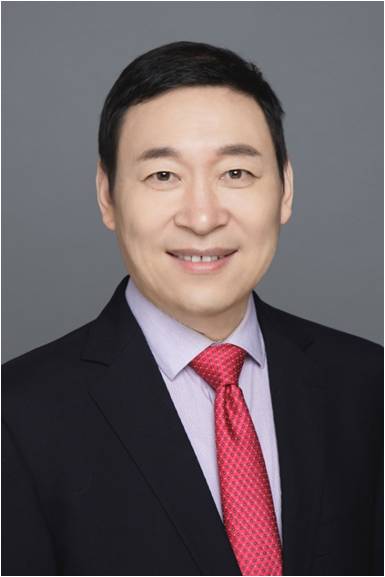
 Lu Hongzhou, Deputy Editor-in-Chief of?Electronic Journal of Emerging Infectious Diseases; Chief Phy...
 -
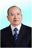
 Yu Weiye, Deputy Editor-in-Chief of?Electronic Journal of Emerging Infectious Diseases; Chief Physic...
 -
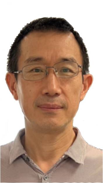
 Yi Yongxiang, Deputy Editor-in-Chief of?Electronic Journal of Emerging Infectious Diseases; Chief Ph...
 -
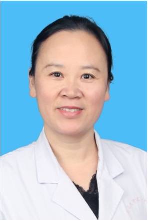
 Zhang Fuchun, Deputy Editor-in-Chief of?Electronic Journal of Emerging Infectious Diseases; Chief Ph...
 -
 Feng Tiejian, Deputy Editor-in-Chief of Electronic Journal of Emerging Infectious Diseases; Chief Te...
 -
 Li Hongjun, Deputy Editor-in-Chief of?Electronic Journal of Emerging Infectious Diseases; Chief Phys...
 -
 Gu Ye, Deputy Editor-in-Chief of?Electronic Journal of Emerging Infectious Diseases; Chief Tradition...
 -
 Lin Jianyan, Deputy Editor-in-Chief of Electronic Journal of Emerging Infectious Diseases; Chief Phy...
 -
 Wang Yixiang, Deputy Editor-in-Chief of?Electronic Journal of Emerging Infectious Diseases; Associat...
 -
 Wu Xiaoping, Deputy Editor-in-Chief of Electronic Journal of Emerging Infectious Diseases; Chief Phy...
 -
 Liu Yuanming, Deputy Editor-in-Chief of Electronic Journal of Emerging Infectious Diseases; Professo...
 -
 Ma Xuejun, Deputy Editor-in-Chief of Electronic Journal of Emerging Infectious Diseases; Director, R...
 -
 Peng Jie, Deputy Editor-in-Chief of Electronic Journal of Emerging Infectious Diseases; Professor, C...
 -
 Shen Yinzhong, Deputy Editor-in-Chief of Electronic Journal of Emerging Infectious Diseases; Chief P...
 -
 Shi Yuxin, Deputy Editor - in - Chief of Electronic Journal of Emerging Infectious Diseases; Chief P...
 -
 Ouyang Xuehui, Executive Editorial Board Member of Electronic Journal of Emerging Infectious Disease...
 -
 Dai Erhei, Executive Editorial Board Member of Electronic Journal of Emerging Infectious Diseases; C...
 -
 Zhou Jie, Executive Editorial Board Member of Electronic Journal of Emerging Infectious Diseases; Ch...
 -
 Jin Guanqiao, Executive Editorial Board Member of Electronic Journal of Emerging Infectious Diseases...
 -
Liu Yubao, Executive Editorial Board Member of Electronic Journal of Emerging Infectious Diseases; C...
-
 Wang Mingmin, Executive Editorial Board Member of Electronic Journal of Emerging Infectious Diseases...
 -
 Li Guimei, Executive Editorial Board Member of Electronic Journal of Emerging Infectious Diseases; S...
 -
 Chen Budong, Editorial Board Member of Electronic Journal of Emerging Infectious Diseases; Chief Phy...
 -
 Liu Jun, Executive Editorial Board Member of Electronic Journal of Emerging Infectious Diseases; Dir...
 -
 Tan Weiguo, Executive Editorial Board Member of Electronic Journal of Emerging Infectious Diseases; ...
 -
 Fang Weijun, Executive Editorial Board Member of Electronic Journal of Emerging Infectious Diseases;...
 -
 Guan Yang, Deputy Editor-in-Chief of Electronic Journal of Emerging Infectious Diseases; Deputy Dire...
 -
 Zhang Ying, Executive Editorial Board Member of Electronic Journal of Emerging Infectious Diseases; ...
 -
 Li Furong, Executive Editorial Board Member of Electronic Journal of Emerging Infectious Diseases; C...
 -
 Xu Chuanjun, Executive Editorial Board Member of Electronic Journal of Emerging Infectious Diseases;...
 -
 Cheng Guanxun, Deputy Editor-in-Chief of Electronic Journal of Emerging Infectious Diseases; Chief P...
 -
 Wang Hui, Executive Editorial Board Member of Electronic Journal of Emerging Infectious Diseases; Di...
 -
 Fang Weijun, Executive Editorial Board Member of Electronic Journal of Emerging Infectious Diseases;...
 -
 Xu Rengen, Executive Editorial Board Member of Electronic Journal of Emerging Infectious Diseases; C...
 -
 Jiang Liangshuang, Executive Editorial Board Member of Electronic Journal of Emerging Infectious Dis...
 -
 Zeng Xianjun, Executive Editorial Board Member of Electronic Journal of Emerging Infectious Diseases...
 -
 Li Tongxia, Executive Editorial Board Member of Electronic Journal of Emerging Infectious Diseases; ...
 -
 Chen Yaokai, Deputy Editor-in-Chief of Electronic Journal of Emerging Infectious Diseases; Professor...
 -
 Zhang Xiaoqin, Executive Editorial Board Member of Electronic Journal of Emerging Infectious Disease...
 -
 Yang Guilin, Executive Editorial Board Member of Electronic Journal of Emerging Infectious Diseases;...
 -
 Yan Xiaofeng, Executive Editorial Board Member of Electronic Journal of Emerging Infectious Diseases...
 -
 Zhang Hong, Executive Editorial Board Member of Electronic Journal of Emerging Infectious Diseases; ...
 -
 He Renliang, Executive Editorial Board Member of Electronic Journal of Emerging Infectious Diseases;...
 -
 Zhang Wenbao, Executive Editorial Board Member of Electronic Journal of Emerging Infectious Diseases...
 -
 Hou Dailun, Executive Editorial Board Member of Electronic Journal of Emerging Infectious Diseases; ...
 -
 Gao Bo, Executive Editorial Board Member of Electronic Journal of Emerging Infectious Diseases; Chie...
 -
 Huang Kui, Executive Editorial Board Member of Electronic Journal of Emerging Infectious Diseases; C...
 -
 He Yulin, Editorial Board Member of Electronic Journal of Emerging Infectious Diseases; Associate Ch...
 -
 Zheng Yongli, Executive Editorial Board Member of Electronic Journal of Emerging Infectious Diseases...
 -
 Hu Guoxin, Chief Physician, Professor; Director of the Department of Infectious Diseases, Peking Uni...
 -
 Yin Yueping, Executive Editorial Board Member of Electronic Journal of Emerging Infectious Diseases;...
 -
 Zhang Yong, Executive Editorial Board Member of Electronic Journal of Emerging Infectious Diseases; ...
 -
 Huang Jian, Executive Editorial Board Member of Electronic Journal of Emerging Infectious Diseases; ...
 -
 Huang Ping, Executive Editorial Board Member of Electronic Journal of Emerging Infectious Diseases; ...
 -
 Zhang Mingxia, Executive Editorial Board Member of Electronic Journal of Emerging Infectious Disease...